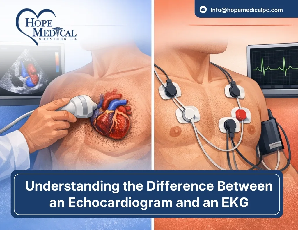
Many people often confuse an echocardiogram (echo) with an electrocardiogram (EKG or ECG), assuming they are the same test because both are related to heart health. However, while both are essential diagnostic tools used by doctors, they serve very different purposes and provide distinct types of information about the heart.
An EKG records the electrical activity of the heart to detect rhythm abnormalities, while an echocardiogram uses ultrasound waves to create detailed images of the heart’s structure and function.
Understanding the differences between these two tests can help patients know what to expect, why a doctor may recommend one over the other, and how each contributes to maintaining heart health.
What is an EKG?
An EKG, also known as an ECG (electrocardiogram), is a quick and painless test that records the electrical activity of your heart. Every time your heart beats, an electrical signal travels through it, triggering the muscle to contract and pump blood. An EKG captures these electrical signals and displays them as waves on a graph.
How Does an EKG Work?
During an EKG, a technician places small, sticky electrodes on your chest, arms, and legs. These electrodes are connected to wires that lead to an EKG machine. The machine detects and records the electrical impulses traveling through your heart with each beat.
The entire process typically takes just 5 to 10 minutes, and you’ll simply lie still while the machine does its work. The resulting printout shows a series of waves and lines that represent different phases of your heartbeat. Your doctor can read this graph to identify abnormalities in your heart’s rhythm, rate, and electrical conduction.
What Can an EKG Detect?
EKGs are particularly useful for diagnosing conditions related to your heart’s electrical system and rhythm, including:
- Arrhythmias: Irregular heartbeats, whether too fast, too slow, or erratic
- Heart attacks: Both current and previous heart attacks leave telltale patterns on an EKG
- Atrial fibrillation: A common irregular heart rhythm
- Heart block: Problems with the electrical signals traveling through your heart
- Coronary artery disease: Reduced blood flow can affect electrical activity
- Heart enlargement: Changes in electrical patterns may suggest an enlarged heart
When Do Doctors Order an EKG?
Your doctor might order an EKG if you’re experiencing symptoms like chest pain, shortness of breath, palpitations, dizziness, or fatigue. It’s also commonly used as a screening tool before surgery, as part of a routine physical for people over 40, or to monitor the effectiveness of heart medications or devices like pacemakers.
What is an Echocardiogram?
An echocardiogram, often called an “echo,” is an ultrasound of your heart. Just like the ultrasound used during pregnancy to see a developing baby, an echocardiogram uses high-frequency sound waves to create moving images of your heart in real-time. This allows doctors to see the actual structure of your heart, how the chambers are moving, and how blood flows through it.
How Does an Echocardiogram Work?
During a standard echocardiogram (transthoracic echo), a technician applies gel to your chest and uses a handheld device called a transducer to capture images. The transducer emits sound waves that bounce off your heart structures and return to create detailed moving pictures on a screen.
You might be asked to lie on your left side or change positions to get different views of your heart.The test usually takes 30 to 60 minutes, and while it’s painless, the technician may need to press firmly with the transducer to get clear images.
Types of Echocardiograms
There are several types of echocardiograms, depending on what your doctor needs to see:
- Transthoracic echocardiogram (TTE): The most common type, performed on the chest
- Transesophageal echocardiogram (TEE): A small probe is inserted down your throat for clearer images
- Stress echocardiogram: Images are taken before and after exercise or medication that makes your heart work harder
- Doppler echocardiogram: Specifically measures blood flow through your heart valves.
What Can an Echocardiogram Detect?
Echocardiograms provide visual information about your heart’s structure and function, making them ideal for detecting:
- Valve problems: Leaking, narrowed, or damaged heart valves
- Heart muscle weakness: Cardiomyopathy or reduced pumping ability
- Congenital heart defects: Structural problems present from birth
- Blood clots or tumors: Masses inside the heart chambers
- Fluid around the heart: Pericardial effusion
- Ejection fraction: How much blood your heart pumps with each beat
- Chamber size: Enlarged or thickened heart chambers
When Do Doctors Order an Echocardiogram?
Your doctor might order an echocardiogram if you have a heart murmur, unexplained shortness of breath, signs of heart failure, chest pain that might be related to heart structure, or if you’ve had a heart attack and they need to assess the damage. It’s also used to monitor known heart conditions and check how well treatments are working.
Differences Between EKG and Echocardiogram
| Feature | EKG (Electrocardiogram) | Echocardiogram |
| Technology Used | Uses electrodes to measure the electrical activity of the heart. Think of it as checking the heart’s wiring. | Uses ultrasound waves to create images of the heart’s structure and motion. Think of it as inspecting the walls, doors, and foundation. |
| What It Measures | Heart rhythm and electrical patterns. Detects irregularities in heartbeat and timing of electrical signals. | Heart structure and function. Shows chamber size, valve function, muscle contraction, and blood flow. |
| Duration and Complexity | Quick and simple, usually 5–10 minutes. | Longer, typically 30–60 minutes, capturing multiple angles and positions. |
| Conditions They Diagnose | Rhythm/electrical issues: arrhythmias, heart attacks, heart block, and coronary artery disease changes. | Structural/mechanical issues: valve disease, heart muscle weakness, congenital defects, fluid accumulation. |
| Cost Considerations | Generally less expensive ($50–$200 without insurance). | More expensive ($300–$2,000 depending on type and location). |
| Preparation Required | No special preparation needed; eat normally and take medications. | Usually, no special preparation; may need fasting fora transesophageal echocardiogram. |
Can EKGs and Echocardiograms Be Done Together?
EKGs and echocardiograms are often performed together because they provide complementary insights into heart health. An EKG detects electrical activity and rhythm abnormalities, such as irregular heartbeats or signs of a heart attack, while an echocardiogram uses ultrasound to evaluate the heart’s structure, pumping ability, and any potential damage or complications.
Having both tests allows doctors to get a comprehensive picture of the heart’s condition. For example, if an EKG shows irregularities, an echocardiogram can determine whether these electrical issues have affected the heart muscle or caused fluid buildup. Together,
They ensure accurate diagnosis and help guide effective treatment planning.
Common Misconceptions About EKGs and Echocardiograms
- One Test is Better Than the Other
Neither test is universally better; an EKG measures electrical activity, while an echocardiogram images heart structure and function. The best test depends on your doctor’s specific clinical question.
- If I Have an Echocardiogram, I Don’t Need an EKG
Having an echocardiogram doesn’t replace an EKG, as they assess different aspects of heart health. Both tests are often useful to get a complete picture.
- These Tests Are Only for People with Heart Problems
Although often used to diagnose heart problems, tests like EKGs and echocardiograms are also useful for routine screening, especially for people over 40, those with risk factors, or athletes monitoring their heart health.
- The Tests Will Hurt
EKGs and standard echocardiograms are painless, non-invasive tests; the only mild discomfort may come from cold gel or removing electrodes.
Conclusion
An EKG and an echocardiogram are two different heart tests, each providing important information. An EKG records the heart’s electrical activity to detect rhythm or signal problems, while an echocardiogram uses ultrasound to visualize the heart’s structure and assess how well its chambers, valves, and muscles function.
Both tests are safe, non-invasive, and help doctors diagnose and manage heart conditions. Knowing the difference helps you understand what to expect—an EKG takes about 10 minutes, while an echocardiogram may take 30 to 60 minutes. Following your doctor’s recommendation for either test is a key step in monitoring and protecting your heart health.
FAQs
Q1. Is an EKG or an echocardiogram more accurate?
Ans: Both are accurate for different purposes; an EKG checks electrical activity, echocardiogram checks heart structure.
Q2. How much does each test cost?
Ans: EKG costs $50–$200, echocardiogram costs $200–$1,000+, depending on type and location.
Q3. Are these tests painful?
Ans: Both are generally painless, with only slight pressure from electrodes or the ultrasound probe.
Q4. Do I need to prepare for either test?
Ans: Minimal preparation is needed; stress echocardiograms may require fasting or avoiding certain meds.
Q5. Can I eat before an echocardiogram or EKG?
Ans: Yes, except for stress echocardiograms, where heavy meals should be avoided.
Q6. Which test is better for detecting heart disease?
Ans: EKG detects rhythm issues; echocardiogram detects structural or valve problems.
Q7. How long does it take to get results?
Ans: EKG results are immediate; echocardiogram results usually take a few hours to a day.