A shortness of breath that lingers after climbing the stairs. Ankles that stay swollen by evening. A fatigue that sleep doesn’t fix. These are the kinds of symptoms that send people searching “can an echo detect heart failure” at midnight, half-hoping the answer is no.
The honest answer is more useful than that: yes, an echocardiogram is one of the most important tools cardiologists use to diagnose heart failure, and it can do far more than confirm a diagnosis. It can explain why the heart is struggling, how severe the problem is, and what kind of treatment will actually help.
How echocardiography fits into a heart failure diagnosis, what a normal versus abnormal echo report looks like, and how an echo test differs from a coronary calcium score, since the two are often confused but measure completely different things.
What Is an Echocardiogram, Exactly?
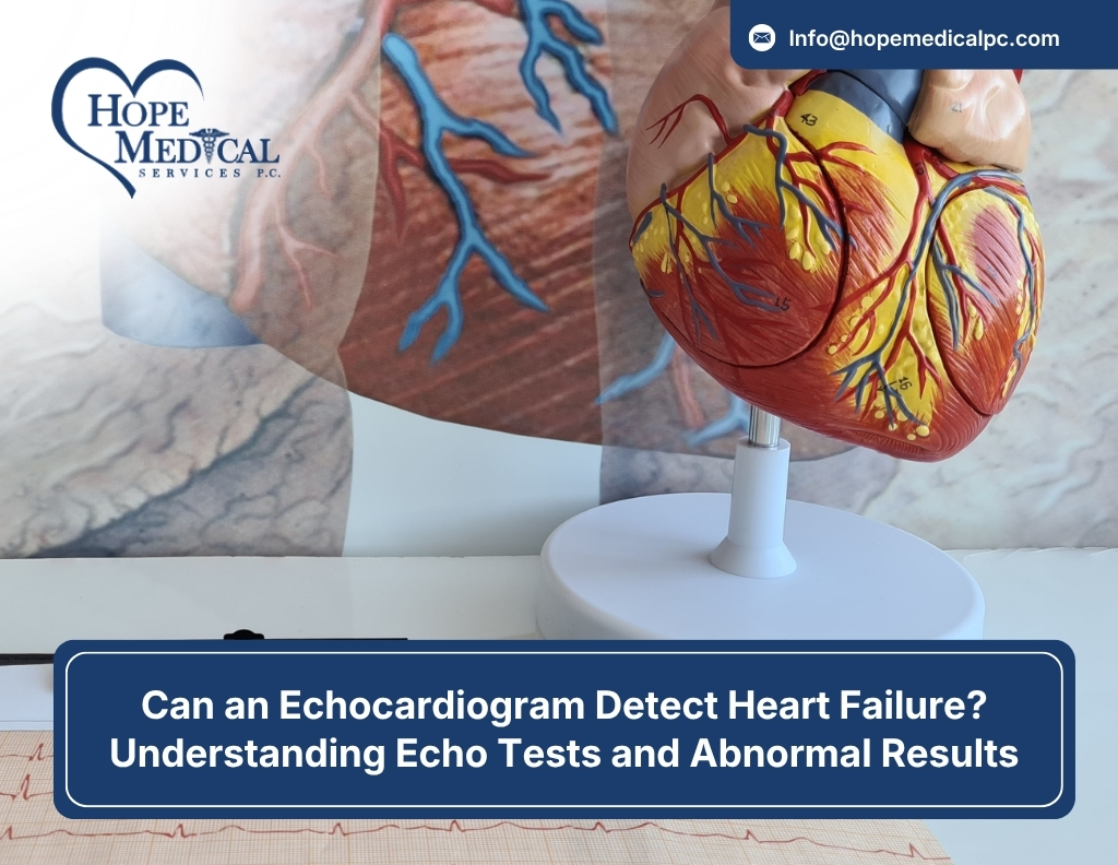
An echocardiogram, or “echo,” is an ultrasound of the heart. A technician moves a small handheld probe across the chest, and sound waves bounce off the heart’s structures to build a live, moving image. There is no radiation, no needles, and for a standard transthoracic echo, no recovery time.
In a single 30- to 45-minute session, an echo can show:
- The size of each heart chamber
- How thick or thin the heart muscle walls are
- How forcefully the heart squeezes with each beat (ejection fraction)
- Whether the four heart valves open and close properly
- Blood flow patterns and direction through the heart
- Fluid buildup around the heart
Can an Echocardiogram Detect Heart Failure?
Yes. Major cardiology guidelines from the American Heart Association and the European Society of Cardiology identify echocardiography as the single most useful test for diagnosing heart failure, because heart failure symptoms alone- breathlessness, swelling, fatigue- are not specific enough to confirm a diagnosis. Anemia, lung disease, and anxiety can all mimic the same symptoms. An echo provides the objective, structural evidence a cardiologist needs to confirm what is actually happening inside the heart.
Rather than relying on symptoms alone, echocardiography in heart failure diagnosis works by measuring specific, quantifiable markers. Here is what the test is actually looking for and why each measurement matters:
| What the Echo Measures | Why It Matters for Heart Failure |
| Ejection Fraction (EF) | The percentage of blood pumped out of the left ventricle with each beat. Low EF points to systolic heart failure. |
| Diastolic Function | How well the heart relaxes and fills with blood between beats. Poor filling suggests diastolic (HFpEF) heart failure. |
| Chamber Size | Enlarged chambers often indicate the heart is straining to compensate for reduced pumping power. |
| Wall Thickness & Motion | Thickened or weakly moving walls can point to prior heart attacks, high blood pressure damage, or cardiomyopathy. |
| Valve Function | Leaking or narrowed valves can both cause and worsen heart failure. |
| Pulmonary Pressures | Elevated pressure in the lungs’ blood vessels is a hallmark sign of a heart that isn’t keeping up. |
How Echocardiography Diagnoses and Stages Heart Failure
Systolic vs. Diastolic Heart Failure on Echo
Heart failure isn’t a single condition; it splits into two distinct patterns, and an echo test for heart function is what tells them apart.
- Heart Failure with Reduced Ejection Fraction (HFrEF): The heart muscle has weakened and can’t squeeze forcefully enough. Echo typically shows an EF below 40%.
- Heart Failure with Preserved Ejection Fraction (HFpEF): The heart squeezes normally but has become stiff and doesn’t relax or fill properly. EF can look normal even though the heart is failing, which is why diastolic measurements on echo are essential, not optional.
This is one of the more misunderstood aspects of heart failure diagnosis: a “normal-looking” ejection fraction number does not automatically rule out heart failure. Nearly half of all heart failure patients have preserved EF, which is exactly why a thorough echo, not just a quick glance at one number, matters so much.
Tracking Heart Failure Progression Over Time
For patients already diagnosed, echocardiography isn’t a one-time event. Cardiologists use repeat echo tests to track whether a treatment plan is working, whether the heart is stabilizing, or whether the disease is advancing. Comparing chamber size, EF, and valve function across visits gives a clearer trend line than any single symptom report can.
Reading Your Echo Test Results: Normal vs. Abnormal Ranges
Once the test is complete, your report will include specific numbers. Here’s a general reference range for the most commonly cited measurement, ejection fraction, so you have context walking into your results discussion. These are general clinical reference points; your cardiologist will interpret your specific numbers in the context of your symptoms and history.
| Ejection Fraction (EF) | General Classification |
| 55% – 70% | Normal heart function |
| 41% – 54% | Mildly reduced / borderline |
| 30% – 40% | Moderately reduced |
| Below 30% | Severely reduced |
A number outside the normal range doesn’t automatically mean a crisis; it means your cardiologist now has objective data to guide the next step, whether that’s medication adjustment, additional imaging, or closer monitoring.
What Does an Abnormal Echocardiogram Mean?
Seeing the word “abnormal” on a report can be unsettling, but it is a broad clinical term, not a diagnosis in itself. An abnormal echocardiogram simply means something on the images fell outside the expected range. The cause can range from minor to significant. Common findings include:
- Mild valve leakage (regurgitation) that many people live with for years without symptoms progressing
- Thickened heart walls from long-standing high blood pressure
- Reduced ejection fraction, suggesting the heart muscle isn’t pumping at full strength
- Enlarged heart chambers, often a sign the heart is compensating for extra strain
- Fluid around the heart (pericardial effusion)
- Signs of a prior, possibly unrecognized, heart attack
When an Abnormal Echo Isn’t an Emergency
Context is everything. A mild finding in someone with no symptoms and a healthy heart history is treated very differently than the same finding in someone with worsening shortness of breath. This is exactly why an abnormal echo should always be followed by a conversation with a cardiologist, rather than an internet search for worst-case scenarios. Your cardiologist may recommend a follow-up echo in a few months simply to watch for change, or order additional testing like an EKG, stress test, or bloodwork to complete the picture.
| Echocardiogram | Calcium Score Test | |
| What it measures | Heart structure, muscle function, valves, and blood flow | Calcium buildup (plaque) in the coronary arteries |
| Technology used | Ultrasound (sound waves) | Low-dose CT scan (X-ray based) |
| Best for detecting | Heart failure, valve disease, cardiomyopathy, enlarged chambers | Coronary artery disease risk and early plaque buildup |
| Typical patient | Someone with symptoms like breathlessness, swelling, or fatigue | Someone assessing long-term heart attack risk, often with no symptoms yet |
| Radiation exposure | None | Low-dose radiation |
An echocardiogram looks at how the heart is functioning right now, while a calcium score estimates future coronary artery disease risk. Many cardiology plans use both tests together, one to evaluate current heart performance, the other to assess long-term risk, because they answer different clinical questions rather than competing with each other.
Signs You May Need an Echo Test
A cardiologist may recommend an echocardiogram if you’re experiencing:
- Shortness of breath, especially during activity or while lying flat
- Swelling in the legs, ankles, or abdomen
- Persistent fatigue that doesn’t improve with rest
- Heart palpitations or an irregular heartbeat
- Chest pain or pressure
- A heart murmur detected during a routine physical exam
- A family history of heart disease or cardiomyopathy
- Follow-up monitoring after a heart attack, stent, or valve procedure
Every Echocardiogram (EKG) test is reviewed directly by a board-certified cardiologist who looks beyond the numbers to understand the full clinical picture, connecting your echo results to your symptoms, history, and long-term heart health plan.
What to Expect During Your Echocardiogram
A standard transthoracic echocardiogram is straightforward and comfortable:
- No fasting or special preparation is typically required
- You’ll lie on an exam table, usually on your left side
- A technician applies gel and moves a probe across your chest
- The test itself takes roughly 30 to 45 minutes
- There’s no downtime; you can resume normal activity immediately after
- Results are reviewed by a cardiologist, who will discuss findings and next steps with you directly
Final Thoughts
An echocardiogram remains one of the fastest, safest, and most informative ways to answer the question that brought you here: can an echo detect heart failure? Yes, and it can also tell your cardiologist exactly what kind of heart failure you’re dealing with, how advanced it is, and which treatment path will help the most. If you’re experiencing symptoms like breathlessness, fatigue, or swelling, don’t wait for them to worsen before getting answers. The team at Hope Medical Services is here to guide you from your first echo test through every step of your heart health journey, with compassionate, precise cardiology care close to home in Queens.
FAQs
Q1. Is an echocardiogram painful?
No. A standard echocardiogram is completely painless and non-invasive. You may feel light pressure from the probe against your chest, but there is no needle, incision, or discomfort involved.
Q2. How long does it take to get echocardiogram results?
In many cases, a cardiologist can review preliminary images the same day. A full formal report with measurements is often available within a few days, and your care team will schedule a follow-up to walk through the findings with you.
Q3. Can an echocardiogram be normal even if I have heart failure symptoms?
Yes, and this is more common than most people realize. Symptoms like fatigue or breathlessness can stem from conditions outside the heart, such as anemia, thyroid issues, or lung disease. If your echo is normal but symptoms persist, your cardiologist will likely order additional testing to find the actual cause.
Q4. Do I need to stop eating or drinking before an echocardiogram?
Typically no. Unlike some cardiac tests, a standard transthoracic echocardiogram usually doesn’t require fasting. Your provider will confirm any specific instructions when your appointment is scheduled.
Q5.How often should someone with heart failure get an echo?
This varies by individual case, but many patients with an existing heart failure diagnosis have a repeat echo roughly once a year, or sooner if symptoms change, medications are adjusted, or a new concern arises.
Q6.Can an echocardiogram detect a heart attack that already happened?
Often, yes. An echo can reveal areas of the heart wall that move weakly or not at all, which frequently indicates scar tissue from a prior, sometimes previously undiagnosed, heart attack.